There was a time when we could only infer the biology of hypertrophic cardiomyopathy (HCM). A patient fainted? Perhaps the myocardium was electrically unstable. The ventricular wall measured 30 mm? The risk must be high. A sibling died suddenly? Better not take chances. These were sensible assumptions, but with increasingly sophisticated clinical tools at our disposal, is that really the best we can do?
Cardiovascular magnetic resonance (CMR) is well-established as the gold standard for tissue characterisation and assessment of myocardial fibrosis in HCM (1). Circulating biomarkers reveal myocardial stress and injury before ventricular dysfunction becomes apparent (2,3). In specific settings, genetics may shed light on why two patients with seemingly comparable phenotypes can follow different clinical trajectories. Yet when one of the most important questions in cardiology arises – should this patient receive an implantable cardioverter-defibrillator (ICD)? – our decision-making still depends predominantly on clinical surrogates developed long before these tools became part of routine practice. When reflecting on the two most widely used risk-stratification criteria for ICD-related decision-making in HCM (Figure 1), are we really using all the tools available in our armamentarium?
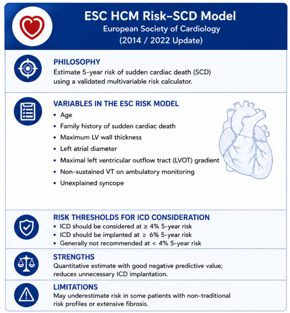
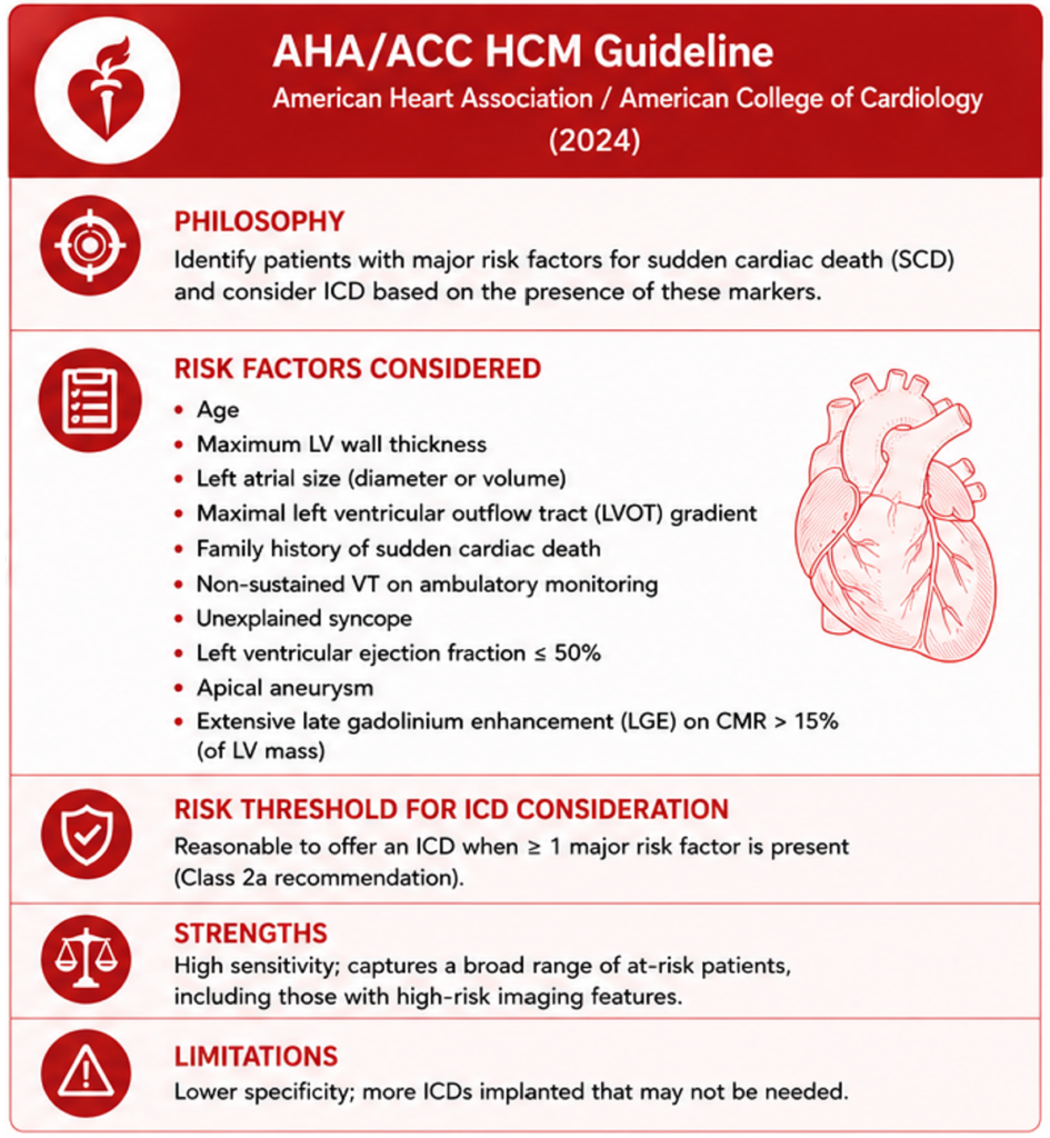
Figure 1. Comparison of current guideline approaches to sudden cardiac death risk stratification in hypertrophic cardiomyopathy (4,5,6).
Consider two patients sitting side by side in the clinic waiting room. Both have HCM. Both have an estimated 5-year sudden cardiac death (SCD) risk of 5% according to the ESC HCM Risk-SCD calculator (Figure 1). Current guidelines would support the same discussion regarding ICD implantation; but their hearts tell very different stories. One has only minimal late gadolinium enhancement (LGE), preserved ventricular architecture and normal NT-proBNP concentrations. The other has extensive myocardial fibrosis,
elevated natriuretic peptides, adverse remodelling and biochemical evidence of ongoing myocardial injury. Although their calculated risk is identical, few cardiologists would genuinely believe that their biological risk is the same. Resolving that disconnect is one of the key challenges of modern HCM care.
Current approaches to risk stratification reflect different compromises. The American Heart Association/American College of Cardiology (AHA/ACC) guidelines prioritise sensitivity, identifying patients with major clinical risk factors who may benefit from an ICD (4). The European Society of Cardiology (ESC) model seeks greater specificity through an integrated risk calculator, reducing unnecessary device implantation (5,6). Neither philosophy is inherently superior; each balances competing risks in a different way. What unites them, however, is that both were developed in an era when we could not routinely measure the myocardial substrate driving ventricular arrhythmias
They also focus almost exclusively on sudden cardiac death (SCD) Although this is the most binary prognostic event an HCM patient can experience, progressive heart failure, atrial fibrillation, stroke, septal reduction-related mortality and the need for advanced heart failure therapies contribute substantially to the burden of disease. Future risk assessment should therefore seek to identify patients at risk of HCM progression, rather than predicting sudden death alone (7).
If there is one lesson that has emerged consistently over the past decade, it is that fibrosis matters. LGE is no longer simply an imaging finding. It represents replacement fibrosis, the very substrate that permits re-entrant ventricular arrhythmias (1). The relationship is biologically plausible, mechanistically coherent and remarkably consistent across observational studies (8,9). The debate is no longer whether fibrosis predicts risk, but how that information should influence clinical decisions.
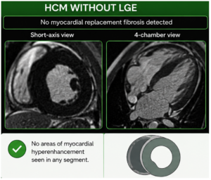
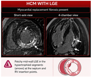
Figure 2. Late gadolinium enhancement (LGE) on cardiovascular magnetic resonance in hypertrophic cardiomyopathy. LGE identifies replacement myocardial fibrosis, an important marker of adverse ventricular remodelling and the arrhythmogenic substrate underlying ventricular arrhythmias and sudden cardiac death (1).
The recently published HCMR registry provides perhaps the clearest evidence yet that the relationship between fibrosis and adverse outcomes is continuous rather than binary (7). Increasing LGE burden was associated with progressively higher risks of major cardiovascular events and SCD-related outcomes, with approximately 9% fibrosis emerging as a potentially discriminatory threshold. These findings align closely with recent meta-analytic evidence suggesting an optimal threshold of around 10%, while simultaneously challenging current guideline definitions of “extensive” fibrosis exceeding 15% (4,9,10).
The same principle was evident elsewhere in HCMR. Measures that directly quantify myocardial disease – LGE burden, left ventricular mass index and indices of adverse remodelling – proved more informative than several traditional clinical variables once contemporary imaging was considered (7). Importantly, these are continuous measurements describing disease burden itself, rather than historical clinical events acting as indirect markers of risk.
Circulating biomarkers add another complementary dimension. Natriuretic peptides reflect myocardial wall stress; high-sensitivity troponin reflects ongoing myocyte injury (2,3).. The integration of imaging findings with biomarkers may provide insight into the current myocardial status, rather than asymmetrically relying on past clinical events to estimate future risk. This represents a fundamental shift in thinking. The real lesson from HCMR is not that we have discovered another risk marker. It is that we may need to rethink what we mean by “risk” in HCM.
Importantly, this is not a call to abandon established risk scores. The ESC calculator remains an easily applicable and rigorously validated tool, while the AHA/ACC framework rightly recognises the importance of clinical features in risk stratification (4,5,6). Ultimately, any risk score is only as good as its users and to be ‘well-used’ it needs to be ‘usable’ in the first place. The argument that adding more parameters would risk cluttering the risk scores and detracting their practical application in a wide range of clinical settings, is therefore a reasonable challenge.
Perhaps the future does not lie in a better risk score at all, but in recognising that no single score can fully capture a disease as biologically heterogeneous as HCM. Therein lies the art of medicine in general, and of HCM-management specifically; rather than rigid algorithms, truly personalised care must leave breathing room for nuance, discourse and deviation. The next generation of HCM risk stratification should aspire to something more ambitious than a better calculator. It should view clinical phenotype, consider multi-parametric radiological and biomarkers, and, ultimately, genotype as complementary pieces of the same puzzle. Such an approach acknowledges an increasingly uncomfortable reality: when the patient in clinic asks about their personal risk, are we truly able to give them a personalised answer? The question is no longer whether we can, but whether our guidelines are ready to catch up?
Reference List:
- Moon et al. (histological basis of LGE) — 10.1016/j.jacc.2004.03.035
- Coats et al. NT-proBNP — 10.1093/eurheartj/eht070
- Kubo et al. hs-cTnT — 10.1016/j.jacc.2013.03.055
- 2024 AHA/ACC Guideline — 10.1016/j.jacc.2024.02.014
- 2023 ESC Cardiomyopathy Guideline — 10.1093/eurheartj/ehad194
- O’Mahony HCM Risk-SCD — 10.1093/eurheartj/eht439
- HCMR Registry (JAMA 2025) — 10.1001/jama.2026.5633
- Chan et al. — 10.1161/CIRCULATIONAHA.113.007094
- Weng et al. meta-analysis — 10.1016/j.jcmg.2016.02.031
- Wang et al. EHJ LGE thresholds — 10.1093/eurheartj/ehad581